
Scoliosis
Definition of Scoliosis
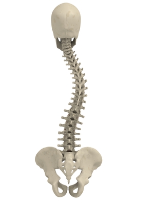
Scoliosis is a medical condition in which a person’s spine is curved from side to side. Although it is a complex three-dimensional deformity, on an X-ray, viewed from the rear, the spine of an individual with scoliosis may look more like an “S” or a “C”, rather than a straight line. Scoliosis is typically classified as either congenital (caused by vertebral anomalies present at birth), idiopathic (cause unknown, subclassified as infantile, juvenile, adolescent, or adult, according to when onset occurred), or neuromuscular (having developed as a secondary symptom of another condition, such as spina bifida, cerebral palsy, spinal muscular atrophy, or physical trauma). A lesser-known cause of scoliosis could be a condition called Chiari malformation.
Recent longitudinal studies reveal that the most common form of the condition, late-onset idiopathic scoliosis, is physiologically harmless and self-limiting. The rarer forms of scoliosis pose risks of complications.
The deformity may begin in the intervertebral discs, producing distortions in the epiphyseal cartilage which may influence the end of growth and therefore the deformity of the vertebrae, resulting in wedging and rotation of the vertebrae.
Cause of Scoliosis
An estimated 65% of scoliosis cases are idiopathic, about 15% are congenital and about 10% are secondary to a neuromuscular disease.
Adolescent idiopathic scoliosis has no clear causal agent, and it is generally believed to be multifactorial, although genetics are believed to play a role. Various causes have been implicated, but none has consensus among scientists as the cause of scoliosis, though the role of genetic factors in its development is widely accepted. Still, at least one gene, notably CHD7, has been associated with the idiopathic form of scoliosis.
Studies in 2006 showed evidence of a linkage between idiopathic scoliosis and three microsatellite polymorphisms in the MATN1 gene (encoding for Matrilin 1, cartilage matrix protein), respectively consisting of 103, 101 and 99 base pairs, .
Congenital scoliosis can be attributed to a malformation of the spine during weeks three to six in utero. It is a result of either a failure of formation, a failure of segmentation, or a combination of stimuli.
Scoliosis secondary to neuromuscular disease may develop during adolescence, such as with tethered spinal cord syndrome. Scoliosis often presents itself, or worsens, during the adolescence growth spurt and is more often diagnosed in females than males.
Signs and Symptoms of Scoliosis
Patients having reached skeletal maturity are less likely to have a worsening case. Some severe cases of scoliosis can lead to diminishing lung capacity, putting pressure on the heart, and restricting physical activities.
The signs of scoliosis can include:
- Uneven musculature on one side of the spine
- A rib prominence and/or a prominent shoulder blade, caused by rotation of the ribcage in thoracic scoliosis
- Uneven hips, arms or leg lengths
- Slow nerve action (in some cases)
Risk Factors for Scoliosis
Scoliosis runs in families. If someone in a family has scoliosis, the likelihood of an incidence is much higher – approximately 20 percent. If anyone in your family has curvature of the spine, you should be examined for scoliosis.
Girls have a much higher risk of the curve worsening and requiring treatment.
Diagnosis of Scoliosis
Scoliosis is defined as a spinal curvature of more than 10 degrees to the right or left as the examiner faces the patient (in the coronal plane). Deformity may also exist to the front or back (in the sagittal plane).
Patients who initially present with scoliosis are examined to determine whether the deformity has an underlying cause. During a physical examination, the following are assessed to exclude the possibility of underlying condition more serious than simple scoliosis
The patient’s gait is assessed, and there is an exam for signs of other abnormalities (e.g., spina bifida as evidenced by a dimple, hairy patch, lipoma, or hemangioma). A thorough neurological examination is also performed, the skin for café au lait spots, indicative of neurofibromatosis, the feet for cavovarus deformity, abdominal reflexes and muscle tone for spasticity.
During the examination, the patient is asked to bend forward as far as possible. This is known as the Adams forward bend test and is often performed on school students. If a prominence is noted, then scoliosis is a possibility and the patient should be sent for an X-ray to confirm the diagnosis.
As an alternative, a scoliometer may be used to diagnose the condition.
When scoliosis is suspected, weight-bearing full-spine AP/coronal (front-back view) and lateral/sagittal (side view) X-rays are usually taken to assess the scoliosis curves and the kyphosis and lordosis, as these can also be affected in individuals with scoliosis. Full-length standing spine X-rays are the standard method for evaluating the severity and progression of the scoliosis, and whether it is congenital or idiopathic in nature.
In growing individuals, serial radiographs are obtained at three- to 12-month intervals to follow curve progression, and, in some instances, MRI investigation is warranted to look at the spinal cord.
The standard method for assessing the curvature quantitatively is measurement of the Cobb angle, which is the angle between two lines, drawn perpendicular to the upper endplate of the uppermost vertebra involved and the lower endplate of the lowest vertebra involved. For patients with two curves, Cobb angles are followed for both curves. In some patients, lateral-bending X-rays are obtained to assess the flexibility of the curves or the primary and compensatory curves.
Genetic testing
Genetic testing for AIS, which became available in 2009 and is still under investigation, attempts to gauge the likelihood of curve progression.
Through a genome-wide association study, geneticists have identified single nucleotide polymorphism markers in the DNA that are significantly associated with adolescent idiopathic scoliosis. Fifty-three genetic markers have been identified. Scoliosis has been described as a biomechanical deformity, the progression of which depends on asymmetric forces otherwise known as the Heuter-Volkmann law.
Prevention of Scoliosis
Scoliosis cannot be prevented. However, measures to increase bone mass and strengthen bones, including getting enough calcium and vitamin D, and doing regular weight-bearing exercise, may help to prevent cases caused by spinal fractures.
If one of your family member has scoliosis, you should have doctor to examine your spine.
Treatment is aimed at preventing the curve from getting worse.
Treatment of Scoliosis
The traditional medical management of scoliosis is complex and is determined by the severity of the curvature and skeletal maturity, which together help predict the likelihood of progression.
The conventional options are, in order:
- Observation
- Physical therapy
- Chiropractic
- Occupational therapy
- Casting (EDF)
- Bracing
- Surgery
A growing body of scientific research testifies to the efficacy of specialized treatment programs of physical therapy, which may include bracing.
Physiotherapy
The Schroth method, a noninvasive, physiotherapeutic treatment, has been used successfully in Europe since the 1920s. The method is based upon the concept of scoliosis as resulting from a complex of muscular asymmetries (especially strength imbalances in the back) that can be at least partially corrected by targeted exercises.
The Schroth method has proven effective at reversing abnormal scoliotic curvatures by an average of 10% in four- to six-week in-patient programs, and by 30% or more in an out-patient program over a period of a year. One study of nearly 200 adolescent Schroth patients found no curve progression three years following the in-patient program. Several studies have documented the Schroth method’s efficacy in substantially reducing or eliminating pain, which tends to be a problem, in particular, for adults.
Small curvatures between 15 and 20° during growth may be treated with the physio-logic-program, curvatures between 20 and 30° during growth spurt with “3D-made-easy”. This program has been tested in the environment of in-patient treatment, as well. In curvatures exceeding 30°, a combination of the methods described together with the Schroth program may be helpful, and a specialized centre with trained and certified staff should be taken into account. Out-patient rehabilitation treatments today may reach the same outcome as in-patient programs. Out-patient programs may be successful when pattern-specific programs are provided. A certain intensity is necessary to allow the very best compliance with conservative treatment, and to acquire strategies for coping with scoliosis and with the conservative treatment.
The indications for treatment depend on degree of curvature, maturity of the patient, and the individual curve pattern. While evidence supporting such conservative, noninvasive treatments is weak, today, conservative management of scoliosis can be regarded as being evidence-based; no substantial evidence has been found to support surgical intervention.
Occupational therapy
An occupational therapist helps those who have experienced an injury or illness regain or maintain the ability to participate in everyday activities. For those with scoliosis, an occupational therapist can provide assistance through assessment, intervention, and ongoing evaluation of the condition. This helps them manage physical symptoms so they can participate in daily activities like self-care, productivity, and leisure.
One intervention involves bracing. During the past several decades, a large variety of bracing devices have been developed for the treatment of scoliosis. Studies demonstrate that preventing force sideways across a joint by bracing prevents further curvature of the spine in idiopathic scoliosis, while other studies have also shown that braces can be used by individuals with scoliosis during physical activities.
Other interventions include postural strategies, such as posture training in sitting, standing, and sleeping positions, and in using positioning supports such as pillows, wedges, rolls, and corsets.
Adaptive and compensatory strategies are also employed to help facilitate individuals to returning daily activities.
Self-care
Disability caused by scoliosis, as well as physical limitations during recovery from treatment-related surgery, often affects an individual’s ability to perform self-care activities. One of the first treatments of scoliosis is the attempt to prevent further curvature of the spine.
Depending on the size of the curvature, this is typically done in one of three ways: bracing, surgery, or postural positioning through customized cushioning. Stopping the progression of the scoliosis can prevent the loss of function in many activities of daily living by maintaining range of motion, preventing deformity of the rib cage, and reducing pain during activities such as bending or lifting.
Occupational therapists are often involved in the process of selection and fabrication of customized cushions. These individualized postural supports are used to maintain the current spinal curvature, or they can be adjusted to assist in the correction of the curvature. This type of treatment can help to maintain mobility for a wheelchair user by preventing the deformity of the rib cage and maintaining an active range of motion in the arms.
For other self-care activities (such as dressing, bathing, grooming, personal hygiene, and feeding), several strategies can be used as a part of occupational therapy treatment.
Environmental adaptations for bathing could include a bath bench, grab bars installed in the shower area, or a handheld shower nozzle. For activities such as dressing and grooming, various assistive devices and strategies can be used to promote independence. An occupational therapist may recommend a long-handled reacher that can be used to assist self-dressing by allowing a person to avoid painful movements such as bending over; a long-handled shoehorn can be used for putting on and removing shoes. Problems with activities such as cutting meat and eating can be addressed by using specialized cutlery, kitchen utensils, or dishes.
Productivity
Productive activities include paid or unpaid work, household chores, school, work, and play. Recent studies in healthcare have led to the development of a variety of treatments to assist in the management of scoliosis thereby maximizing productivity for people of all ages.
Assistive technology has undergone dramatic changes over the past 20 years; the availability and quality of the technology has improved greatly. As a result of using assistive technology, functional changes may range from improvements in abilities, performance in daily activities, participation levels, and quality of life.
A common assistive technology intervention is specialized seating and postural control. For children with poor postural control, a comfortable seating system that provides them with the support needed to maintain a sitting position can be essential for raising their overall level of well-being. A child’s well-being in a productive sense involves the ability to participate in classroom and play activities. Specialized wheelchair seating has been identified as the most common prescription in the management of scoliosis in teenagers with muscular dystrophy.
With comfortable wheelchair seating, teenagers are able to participate in classroom activities for longer periods with less fatigue. By tilting the seating position 20° forward (toward the thighs), seating pressure is significantly redistributed, so sitting is more comfortable. If an office worker with scoliosis can sit for longer periods, increased work output is likely to occur and could improve quality of life. Tall, forward-sloping seats or front parts of seats, and when possible with tall desk with the opposite slope, can, in general, reduce pains and the need of bending significantly while working or studying, and that is particularly important with braced, fragile, or tender backs. An open hip angle can benefit the used lung volume and respiration.
For those not using a wheelchair, bracing may be used to treat scoliosis. Lifestyle changes are made to compensate for the proper use of spine braces.
Leisure
Many physical symptoms can prevent a person from engaging in physical leisurely activities, such as chest pains, back pains, shortness of breath, and limited spinal movement. The occupational therapist’s role is to help individuals with scoliosis manage these physical symptoms so they can participate in physical leisure activities.
Bracing is a common strategy recommended by an occupational therapist, in particular, for individuals engaging in sports and exercise. An OT is responsible for educating an individual on the advantages and disadvantages of different braces, proper ways to wear the brace, and the day-to-day care of the brace.
To help a person manage heart and lung symptoms, such as shortness of breath or chest pains, an occupational therapist can teach the individual energy conservation techniques. This includes scheduling routine breaks during the activity, as suitable for the individual. For example, an occupational therapist can recommend that a swimmer take breaks between laps to conserve energy. Adapting or modifying the exercise or sport is another way a person with scoliosis can do it. Adapting the activity may change the difficulty of the sport or exercise. For example, it might mean taking breaks throughout an exercise. If a person with scoliosis is unable to participate in a sport or exercise, an OT can help the individual explore other physical activities that are suitable to his/her interests and capabilities. An occupational therapist and the person with scoliosis can explore enjoyable and meaningful participation in the sport/exercise in another capacity, such as coaching or refereeing.
Bracing
Bracing is normally done when the patient has bone growth remaining and is, in general, implemented to hold the curve and prevent it from progressing to the point where surgery is recommended. In some cases with juveniles, bracing has reduced curves significantly, going from a 40 degrees (of the curve, mentioned in length above.) out of the brace to 18 degrees in it. Braces are sometimes prescribed for adults to relieve pain related to scoliosis.
Bracing involves fitting the patient with a device that covers the torso; in some cases, it extends to the neck. The most commonly used brace is a TLSO, such as a Boston brace, a corset-like appliance that fits from armpits to hips and is custom-made from fiberglass or plastic. It is sometimes worn 22–23 hours a day, depending on the doctor’s prescription, and applies pressure on the curves in the spine. The effectiveness of the brace depends not only on brace design and orthotist skill but on patient compliance and amount of wear per day.
The typical use of braces is for idiopathic curves that are not grave enough to warrant surgery, but they may also be used to prevent the progression of more severe curves in young children, to buy the child time to grow before performing surgery, which would prevent further growth in the part of the spine affected.
Casting
In progressive infantile and sometimes juvenile scoliosis, a plaster jacket applied early may be used instead of a brace. It has been proven possible to permanently correct cases of infantile idiopathic scoliosis by applying a series of plaster casts (EDF: elongation, derotation, flexion) on a specialized frame under corrective traction, which helps to “mould” the infant’s soft bones and work with their growth spurts. This method was pioneered by UK scoliosis specialist Min Mehta. EDF casting is now the only clinically known nonsurgical method of complete correction in progressive infantile scoliosis. Complete correction may be obtained for curves less than 50° if the treatment begins before the second year of life.
Surgery
Surgery is usually recommended by orthopedists for curves with a high likelihood of progression (i.e., greater than 45 to 50° of magnitude), curves that would be cosmetically unacceptable as an adult, curves in patients with spina bifida and cerebral palsy that interfere with sitting and care, and curves that affect physiological functions such as breathing.
Surgery for scoliosis is performed by a surgeon specializing in spine surgery. For various reasons, it is usually impossible to completely straighten a scoliotic spine, but in most cases, significant corrections are achieved.
The two main types of surgery are:
- Anterior fusion: This surgical approach is through an incision at the side of the chest wall.
- Posterior fusion: This surgical approach is through an incision on the back and involves the use of metal instrumentation to correct the curve.
One or both of these surgical procedures may be needed. The surgery may be done in one or two stages and, on average, takes four to eight hours.
Spinal fusion with instrumentation
Spinal fusion is the most widely performed surgery for scoliosis. In this procedure, bone is grafted to the vertebrae so when they heal, they form one solid bone mass and the vertebral column becomes rigid. This prevents worsening of the curve, at the expense of some spinal movement. This can be performed from the anterior (front) aspect of the spine by entering the thoracic or abdominal cavities, or more commonly, performed from the back (posterior). A combination is used in more severe cases.
Spinal fusions were once performed without metal implants. In this technique, a cast was applied after the surgery, usually under traction, to pull the curve as straight as possible, and then hold it there while fusion took place. Casting left patients largely immobilized for a period of weeks to months, with significant burden on patient quality of life. Additionally, the risk of pseudarthrosis (fusion failure) at one or more levels was relatively high, and significant correction could not always be achieved.
Modern spinal systems are attempting to address sagittal imbalance and rotational defects unresolved by the Harrington rod system. They involve a combination of rods, screws, hooks, and wires fixing the spine, and can apply stronger, safer forces to the spine than the Harrington rod. This technique, the Cotrel-Dubousset instrumentation, is currently the most common technique for the procedure.
In general, modern spinal fusions have good outcomes with high degrees of correction and low rates of failure and infection. Patients with fused spines and permanent implants tend to have normal lives with unrestricted activities when they are younger; it remains to be seen whether those that have been treated with the newer surgical techniques develop problems as they age.
Pedicle screw-only posterior spinal fusion may improve major curve correction at two years among patients with adolescent idiopathic scoliosis (AIS) as compared to hybrid instrumentation (proximal hooks with distal pedicle screws) (65% versus 46%) according to a retrospective, matched-cohort study. The prospective cohorts were matched to the retrospective cohorts according to patient age, fusion levels, Lenke curve type, and operative method. The two groups were not significantly different in regard to age, Lenke AIS curve type, or Riser grade. The numbers of fused vertebrae were significantly different (11.7±1.6 for pedicle screw versus 13.0±1.2 for hybrid group). This study’s results may be biased due to the pedicle screw group’s being analyzed prospectively versus retrospective analysis of the hybrid instrumentation group.
Thoracoplasty
A complementary surgical procedure a surgeon may recommend is called thoracoplasty (also called costoplasty). This is a procedure to reduce the rib hump that affects most scoliosis patients with a thoracic curve. A rib hump is evidence of some rotational deformity to the spine.
Thoracoplasty may also be performed to obtain bone grafts from the ribs instead of the pelvis, regardless of whether a rib hump is present. Thoracoplasty can be performed as part of a spinal fusion or as a separate surgery, entirely.
Thoracoplasty is the removal (or resection) of typically four to six segments of adjacent ribs that protrude. Each segment is one to two inches long. The surgeon decides which ribs to resect based on either their prominence or their likelihood to be realigned by correction of the curvature alone. The ribs grow back straight.
Thoracoplasty has risks, such as increased pain in the rib area during recovery or reduced pulmonary function (10–15% is typical) following surgery. This impairment can last anywhere from a few months to two years. Because thoracoplasty may lengthen the duration of surgery, patients may also lose more blood or develop complications from the prolonged anesthesia. A more significant, though far less common, risk is the surgeon might inadvertently puncture the pleura, a protective coating over the lungs. This could cause blood or air to drain into the chest cavity, haemothorax or pneumothorax, respectively.
Complications
The risk of undergoing surgery for scoliosis is estimated to be 5%. Possible complications may be inflammation of the soft tissue or deep inflammatory processes, breathing impairments, bleeding and nerve injuries. However, according to the latest evidence, the rate of complications is far higher. As early as five years after surgery, another 5% require reoperation, and today it is not yet clear what to expect from spine surgery in the long term.
Taking into account that signs and symptoms of spinal deformity cannot be changed by surgical intervention, surgery remains primarily a cosmetic indication, only especially in patients with adolescent idiopathic scoliosis, the most common form of scoliosis never exceeding 80°.
However, the cosmetic effects of surgery are not necessarily stable.
If one decides to undergo surgery, a specialized center should be preferred.
Surgery without fusion
New implants that aim to delay spinal fusion and to allow more spinal growth in young children have been developed. For the youngest patients, whose thoracic insufficiency compromises their ability to breathe and applies significant cardiac pressure, ribcage implants that push the ribs apart on the concave side of the curve may be especially useful.
These Vertical, Expandable Prosthetic Titanium Ribs (VEPTR) provide the benefit of expanding the thoracic cavity and straightening the spine in all three dimensions while allowing it to grow.
An expandable rod, called a growing rod, may be surgically implanted across the segment of spinal curvature, and lengthened, under surgery, every six months to mimic and maintain normal spine growth. This intervention can halt the progress of curvature and gradually straighten the spine. A magnetically controlled growing rod (MCGR) system has been developed and is undergoing clinical trials in Hong Kong. This intervention employs a rod that may be expanded by applying strong magnets to the outside of the patient’s body, and so does not involve repeated surgeries. The recently published report on the first two children to reach 24 month follow-up (aged 5 and 12 years) is encouraging.
Monthly rod lengthening produced a progressive increase in spinal length and correction of scoliosis was maintained.
Although these methods are novel and promising, they are suitable only for growing patients. Spinal fusion remains the “gold standard” of surgical treatment for scoliosis.